

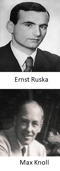
The Transmission electron microscope (TEM) is a special type of microscope that uses electrons to create high magnification images of the internal structure of a sample. Ernst Ruska designed and developed the TEM during his graduate studies under the supervision of Max Knoll in 1932-1933. However, the first commercial electron microscope was installed in IG Farben-Werke in 1939.
Ernst Ruska received the 1986 Nobel Prize in physics for his fundamental work in electron optics and for the design of the first electron microscope. Honestly, it will surprise you that the first electron microscope was less powerful than its optical microscope.
 |  |
What is the difference between TEM and optical microscope?
Abbe equation (1873) : Resolution of a microscope
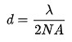
d = resolution λ = wavelength
NA = Numerical aperture
De Broglie equation (1924): Electrons have both waves and particles like properties

λ = wavelength
h = Planck’s constant (6.6 X 10⁻²⁷)
m = mass of the particle (9.1 X 10⁻²⁸ g) v = velocity of the particle
| Microscope | Resolution | Magnification | Lens | Light Source |
| Optical | 200 nm | 1000x | Optical | Visible or UV light |
| TEM | 0.2 nm | 500000x | Electromagnetic | Electron |
•The TEM operates on the same basic principles as the light microscope
•In a TEM, electrons replace photons, electromagnetic lenses replace glass lenses, and images are viewed on a screen rather than through an eyepiece or CCD camera.
• TEM uses the wave nature of electrons
•Electrons create images based on the transmission of the electrons through the sample
What constitutes a TEM?
| •Electron Source •Electromagnetic Lens •Specimen stage •Imaging System •Vacuum system |  |
The electron source consists of a cathode and an anode
Tungsten filament or a lanthanum hexaboride (LaB6) single crystal acts as emission source or cathode
The gun is connected to a high voltage source (typically 80–300 kV)
The beam is then accelerated towards the specimen by the positive anode
Electrons at the rim of the beam will fall onto the anode while the others at the center will pass through the small hole of the anode.
The electron beam is tightly focused using electromagnetic lens and metal apertures
Circular electro-magnets capable of generating a precise circular magnetic field. The field acts like an optical lens to focus the electrons
Aperture is used to restrict the electron beam and filter out unwanted electrons before hitting the specimen. It is a thin disk with a small (2-100 micrometers) circular through-hole
The electromagnetic lens system only allows electrons within a small energy range to pass through, so the electrons in the electron beam will have a well-defined energy
Vacuum system allows the voltage difference between the cathode and the ground without generating an arc
Vacuum system also reduce the collision frequency of electrons with gas atoms to negligible levels
TEMs are equipped with multiple pumping systems and airlocks and are not permanently vacuum sealed
Low vacuum pump: Rotary vane pump or diaphragm (10−4 Pa)
High vacuum pump: turbo-molecular or diffusion (10−4 to 10−7 Pa)
Ultra-high vacuums pumps: Ion pumps (10−7 to 10−9 Pa)
TEM specimen stage includes several airlocks to allow the insertion of the specimen holder into the vacuum with minimal loss of vacuum inside the TEM
A TEM stage hold a specimen and bring the region of interest of the specimen into the path of the electron beam
In the side entry Specimen stage the specimen holder is placed near the tip of a long metal rod, with the specimen placed flat in a small bore with the help of a metal grid
Grid Materials: Nickel, copper, molybdenum, gold or platinum
TEM grid sizes: Diameter 3.05 mm
The electron image is monochromatic
The image is made visible to the eye by allowing the electrons to fall on a fluorescent screen fitted at the base of the microscope column
In digital imaging system the image is captured on a CCD camera and displayed on a computer monitor
What is the easiest way of preparing a TEM sample
TEM specimens should be less than 100 nanometers thick
Preparation of TEM specimens is specific to the material and the type of information to be obtained from the specimen
Nanomaterials and small organisms can be prepared by dipping the grids into a dilute sample containing the specimen.
Biological specimens is embedded in resin that enables cutting of tissue into electron transparent thin sections
What is the working principle of TEM?
•A coherent beam of electron is focused by the use of the condenser lens
•High angle electrons are removed by inserting the condenser aperture
•The electron beam strikes the sample and parts of it are transmitted through it
•Objective lens then focused this transmitted beam to the viewing screen
•While passing the column through the projector lenses the image become enlarged
• On the fluorescent screen or charge coupled device (CCD) camera the users can see the image
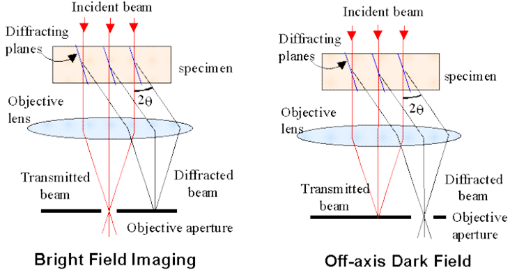
Imaging in TEM
Bright Field imaging and Dark Field imaging
In Bright Field imaging mode, objective aperture only allows the transmitted electron to pass through the electromagnetic lens
In Dark Field imaging mode, objective aperture only allow the diffracted beam to pass through the electromagnetic lens
TEM for special purposes
Scanning TEM (STEM): Rasters the beam across the sample to form the image
Cryo-TEM: Uses a TEM with a specimen holder capable of maintaining the specimen at liquid nitrogen or liquid helium temperatures. Suitable for imaging of materials that are volatile in high vacuum at room temperature and biological sample
Environmental/In-situ TEM: In-situ experiments may also be conducted in TEM using differentially pumped sample chambers, or specialized holders
Aberration Corrected TEM: Modern TEM where aberration of the lens are corrected to reduce the amount of distortion in the image
Low-voltage electron microscope: Voltage used between 5–25 kV and important for biological specimens
Most common uses of TEM
•Nanotechnology
•Biological research
•Medical research
•Material research
•Forensic analysis
•Gemology
•Metallurgy
What are the advantages of TEM?
•TEM offer the most powerful magnification
•Images are high-quality and detailed
•TEM have a wide-range of applications in scientific, educational and industrial fields
•TEM provide information on element and compound structure
•TEM are able to yield information of surface features, shape, size and structure
•TEM are easy to operate with proper training
What are the disadvantages of TEM?
•TEM are large and very expensive
•Laborious sample preparation
•Potential artifacts from sample preparation
•Operation and analysis requires special training
•Samples are limited to those that are electron transparent, able to tolerate the vacuum chamber and small enough to fit in the chamber
•TEM require special housing and maintenance
•Images are black and white